

EYE (VISION)
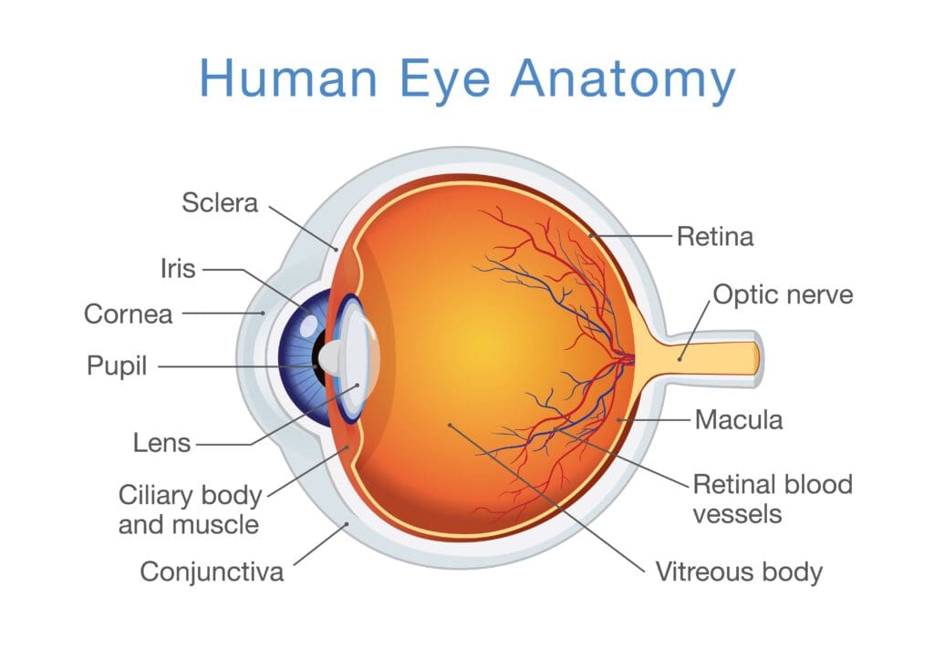
The basic structure of the eye is similar to a simple camera with an aperture (the iris), a lens, and a light sensitive surface (the retina). Light enters the eye through the cornea, then passes through the iris and the lens and falls on the retina. Here the light stimulates the light-sensitive cells on the retina (rods and cones) and these pass small electrical impulses by way of the optic nerve to the visual cortex in the brain. Here, the electrical impulses are interpreted and an image is perceived.
The Cornea:
The cornea is a clear ‘window’ at the very front of the eye. The cornea acts as a fixed focusing device. The focusing is achieved by the shape of the cornea bending the incoming light rays. The cornea is responsible for between 70% and 80% of the total focusing ability (refraction) of the eye.
The Iris and Pupil:
The iris (the coloured part of the eye) controls the amount of light that is allowed to enter the eye. It does this by varying the size of the pupil (the dark area in the centre of the iris). The size of the pupil can be changed very rapidly to cater for changing light levels. The amount of light can be adjusted by a factor of 5:1.
The Lens:
After passing through the pupil, the light passes through the lens. Its shape is changed by the muscles (cillary muscles) surrounding it which results in the final focusing adjustment to place a sharp image onto the retina. The change of shape of the lens is called accommodation. In order to focus clearly on a near object, the lens is thickened. To focus on a distant point, the lens is flattened. The degree of accommodation can be affected by factors such as fatigue or the ageing process.
visual acuity:
When a person is tired accommodation is reduced, resulting in less sharp vision (sharpness of vision is known as visual acuity).Visual acuity is the ability of the eye to discriminate sharp detail at varying distances.
The Retina:
The retina is located on the rear wall of the eyeball. It is made up of a complex layer of nerve cells connected to the optic nerve. Two types of light sensitive cells are found in the retina - rods and cones. The central area of the retina is known as the fovea and the receptors in this area are all cones. It is here that the visual image is typically focused. Moving outwards, the cones become less dense and are progressively replaced by rods, so that in the periphery of the retina, there are only rods.
Cones function in good light and are capable of detecting fine detail and are colour sensitive. This means the human eye can distinguish about 1000 different shades of colour.
Rods cannot detect colour. They are poor at distinguishing fine detail, but good at detecting movement in the edge of the visual field (peripheral vision). They are much more sensitive at lower light levels. As light decreases, the sensing task is passed from the cones to the rods. This means in poor light levels we see only in black and white and shades of grey.
WHY EYE IS MORE SENSITIVE THAN EAR ?
The eye is very sensitive in the right conditions (e.g. clear air, good light, etc.). In fact, the eye has approximately 1.2 million nerve cells leading from the retinas to the area of the brain responsible for vision, while there are only about 50,000 from the inner ears - making the eye about 24 times more sensitive than the ear.
Various factors can affect and limit the visual acuity of the eye.
These include:
• Physical factors such as: • physical imperfections in one or both eyes (short sightedness, long sightedness), • age. • The influence of ingested foreign substances such as: • drugs, • medication, • alcohol, • cigarettes.
• Environmental factors such as:
• amount of light available, • clarity of the air (e.g. dust, mist, rain, etc.). • Factors associated with object being viewed such as: • size and contours of the object, • contrast of the object with its surroundings, • relative motion of the object, • distance of the object from the viewer, • the angle of the object from the viewer.
Hearing
The Basic Function of the Ear:
The ear performs two quite different functions. It is used to detect sounds by receiving vibrations in the air, and secondly, it is responsible for balance and sensing acceleration. Of these two, the hearing aspect is more pertinent to the maintenance engineer, and thus it is necessary to have a basic appreciation of how the ear works.
: outer ear, middle ear and inner ear. These act to receive vibrations from the air and turn these signals into nerve impulses that the brain can recognise as sounds.

Outer Ear:
The outer part of the ear directs sounds down the auditory canal, and on to the eardrum. The sound waves will cause the eardrum to vibrate.
Middle Ear:
Beyond the eardrum is the middle ear which transmits vibrations from the eardrum by way of three small bones known as the ossicles, to the fluid of the inner ear. The middle ear also contains two muscles which help to protect the ear from sounds above 80 dB by means of the acoustic or aural reflex, reducing the noise level by up to 20 dB. However, this protection can only be provided for a maximum of about 15 minutes, and does not provide protection against sudden impulse noise such as gunfire. It does explain why a person is temporarily ‘deafened’ for a few seconds after a sudden loud noise. The middle ear is usually filled with air which is refreshed by way of the eustachian tube which connects this part of the ear with the back of the nose and mouth. However, this tube can allow mucus to travel to the middle ear which can build up, interfering with normal hearing.
Inner Ear:
Unlike the middle ear, the inner ear is filled with fluid. The last of the ossicles in the middle ear is connected to the cochlea. This contains a fine membrane (the basilar membrane) covered in hair-like cells which are sensitive to movement in the fluid. Any vibrations they detect cause neural impulses to be transmitted to the brain via the auditory nerve.

New
You must be logged in to post a comment.