
Introduction
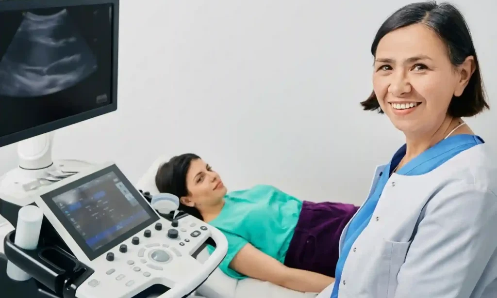
Modern medical imaging has changed the way doctors diagnose and monitor many health conditions. 3D ultrasound imaging creates detailed, three-dimensional views that help healthcare providers examine organs, tissues, and developing babies with greater clarity than traditional two-dimensional ultrasound. This advanced technology is commonly used during pregnancy, but it also supports the evaluation of various medical conditions involving muscles, blood vessels, and internal organs. At ER of Fort Worth, patients have access to advanced diagnostic technology and experienced emergency medical professionals who are committed to providing fast, accurate, and compassionate care whenever it is needed.
What Is 3D Ultrasound Imaging?
3D ultrasound imaging is an advanced form of ultrasound that combines multiple two-dimensional images to create a three-dimensional picture. Unlike standard ultrasound, which displays flat images, this technology provides depth and greater detail.
Doctors use sound waves instead of radiation, making ultrasound one of the safest imaging methods available for many patients.
This imaging technology helps healthcare providers:
- View organs from different angles
- Examine soft tissues in greater detail
- Monitor fetal growth during pregnancy
- Evaluate certain injuries
- Assist in diagnosing various medical conditions
How Does 3D Ultrasound Work?
Ultrasound uses high-frequency sound waves that travel through the body. These sound waves bounce back after hitting tissues and organs. A computer processes these signals and creates images.
With 3D ultrasound, multiple image slices are combined into one detailed three-dimensional image.
The process usually includes:
- Applying ultrasound gel to the skin
- Moving a handheld transducer across the area
- Capturing multiple images
- Creating a detailed 3D view within seconds
The examination is painless and usually takes between 20 and 45 minutes depending on the area being examined.
Common Uses of 3D Ultrasound Imaging
Although many people associate 3D ultrasound with pregnancy, it has many medical applications.
Pregnancy Monitoring
One of the most common uses is prenatal care. Doctors may recommend 3D ultrasound to:
- Monitor fetal development
- Check facial structures
- Detect certain birth abnormalities
- Evaluate fetal movement
- Improve visualization when needed
It can provide parents with clearer images while also helping physicians make more accurate assessments.
Gynecological Evaluation
Healthcare providers use 3D ultrasound to evaluate:
- Uterus
- Ovaries
- Fibroids
- Ovarian cysts
- Congenital uterine abnormalities
Detailed imaging often improves diagnostic accuracy.
Heart and Blood Vessel Assessment
Ultrasound technology helps evaluate:
- Blood flow
- Heart valves
- Blood vessel abnormalities
- Circulation problems
These examinations help physicians identify cardiovascular concerns without using radiation.
Musculoskeletal Imaging
Doctors may use ultrasound to examine:
- Tendons
- Ligaments
- Muscles
- Soft tissue injuries
- Joint inflammation
This can assist in diagnosing sports injuries and other musculoskeletal conditions.
Abdominal Organ Evaluation
3D ultrasound may also help examine:
- Liver
- Gallbladder
- Kidneys
- Pancreas
- Spleen
- Bladder
It assists physicians in identifying abnormalities that may require additional evaluation or treatment.
Benefits of 3D Ultrasound Imaging
This advanced technology offers several important advantages.
Better Image Quality
Three-dimensional images provide improved visualization of anatomy, making certain conditions easier to evaluate.
No Radiation Exposure
Unlike CT scans or X-rays, ultrasound uses sound waves instead of radiation.
This makes it a preferred imaging option in many situations, especially during pregnancy.
Non-Invasive Procedure
No needles, incisions, or anesthesia are required for most ultrasound examinations.
Patients can usually return to normal activities immediately after the scan.
Fast Results
Many ultrasound examinations can be completed quickly, allowing physicians to review images without lengthy delays.
Improved Diagnostic Accuracy
Detailed imaging helps physicians evaluate structures more completely, supporting more informed medical decisions.
Who May Need 3D Ultrasound Imaging?
A healthcare provider may recommend this imaging for patients who have:
- Pregnancy evaluations
- Pelvic pain
- Abdominal pain
- Soft tissue injuries
- Suspected organ abnormalities
- Blood vessel concerns
- Follow-up imaging after previous tests
The decision depends on individual symptoms and medical history.
What to Expect During Your Appointment
Patients often wonder what happens during a 3D ultrasound examination.
Typically, the process includes:
- Patient registration
- Medical history review
- Positioning on the examination table
- Application of ultrasound gel
- Image collection
- Image review by a physician
The procedure is generally comfortable and requires little preparation.
Is 3D Ultrasound Safe?
Yes. Ultrasound has been used safely in medicine for many decades.
Since it uses sound waves instead of radiation, it is considered a safe diagnostic tool when performed by trained healthcare professionals.
However, ultrasounds should always be performed for medical purposes under professional supervision.
3D Ultrasound vs Traditional 2D Ultrasound
| Feature | 2D Ultrasound | 3D Ultrasound |
|---|---|---|
| Image Type | Flat image | Three-dimensional image |
| Detail Level | Standard | Higher detail |
| Pregnancy Images | Basic | More realistic |
| Organ Visualization | Good | Enhanced |
| Radiation | None | None |
Both technologies have important roles depending on the patient's condition.
When Should You Visit the Emergency Room?
While many ultrasounds are scheduled appointments, some symptoms require immediate medical attention.
Visit ER of Fort Worth immediately if you experience:
- Severe abdominal pain
- Heavy bleeding during pregnancy
- Sudden pelvic pain
- Serious injury
- Severe swelling
- Difficulty breathing
- Chest pain
- Signs of internal bleeding
Prompt evaluation can help identify serious conditions quickly.
Why Choose ER of Fort Worth?
ER of Fort Worth is committed to providing high-quality emergency medical care with advanced diagnostic capabilities.
Patients benefit from:
- Advanced imaging technology
- Experienced emergency physicians
- Fast medical evaluations
- Minimal waiting times
- Compassionate patient care
- Comprehensive emergency services
- Modern medical equipment
- Care available 24/7
Whether you need emergency evaluation or advanced diagnostic imaging, the team is dedicated to delivering timely and accurate care.
Tips Before Your Ultrasound
To help ensure a successful examination:
- Follow your doctor's preparation instructions.
- Wear comfortable clothing.
- Bring previous imaging reports if available.
- Stay hydrated if instructed.
- Arrive a few minutes early.
- Inform your healthcare provider about any medical conditions.
Conclusion
3D ultrasound has become an important part of modern healthcare by providing detailed, real-time images that help physicians diagnose and monitor many medical conditions. From pregnancy evaluations to abdominal and musculoskeletal assessments, this technology supports accurate medical decision-making while avoiding radiation exposure. When urgent symptoms require immediate attention,ER of Fort Worth offers advanced diagnostic services, experienced emergency professionals, and compassionate care to help patients receive timely treatment.
For more information visit:https://eroffortworthtx.com/services/3d-ultrasound-fort-worth/

You must be logged in to post a comment.