
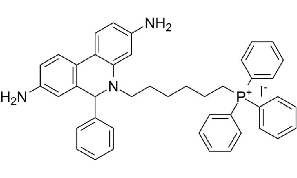
MitoSOX Red is a live cell fluorescent probe targeting mitochondria with maximum excitation/emission light of 510/580 nm.
Products are for research use only. Not for human use. We do not sell to patients.
MitoSOX Red is a live cell fluorescent probe targeting mitochondria with a maximum excitation/emission light of 510/580 nm. MitoSOX Red has cell membrane permeability and can quickly and selectively target mitochondria after entering cells. After entering mitochondria, MitoSOX Red is easily oxidized by superoxide and produces strong red fluorescence after combining with nucleic acids in mitochondria. MitoSOX Red can be used as a fluorescent indicator to specifically detect superoxide[1-2].
Dihydroethidium, also called hydroethidine (HE), can be oxidized by reactive species and subsequently binds to DNA to produce fluorescence. MitoSOX Red is a dihydroethidium derivative with a cationic triphenylphosphonium moiety. This positively charged probe rapidly accumulates in mitochondria and can therefore be used to monitor superoxide/ROS production within mitochondria by fluorometry, microscopy, or flow cytometry[3]. For example, in NK cells, MitoSOX red can observe the distribution of mitochondrial ROS[1]. Confocal microscopy imaging shows that in H9c2 cells treated with paraquat, the fluorescence intensity of MitoSOX Red-labeled mitochondria increases significantly[4].
MitoSOX Red
References:
[1] Jin F, Wu Z, Hu X, Zhang J, Gao Z, Han X, Qin J, Li C, Wang Y. The PI3K/Akt/GSK-3β/ROS/eIF2B pathway promotes breast cancer growth and metastasis via suppression of NK cell cytotoxicity and tumor cell susceptibility. Cancer Biol Med. 2019 Feb;16(1):38-54.
[2] Milliken A S, Nadtochiy S M, Brookes P S. Inhibiting succinate release worsens cardiac reperfusion injury by enhancing mitochondrial reactive oxygen species generation[J]. Journal of the American Heart Association, 2022, 11(13): e026135.
[3] Kauffman ME, Kauffman MK, Traore K, Zhu H, Trush MA, Jia Z, Li YR. MitoSOX-Based Flow Cytometry for Detecting Mitochondrial ROS. React Oxyg Species (Apex). 2016;2(5):361-370.
[4] Mukhopadhyay P, Rajesh M, Yoshihiro K, et al. Simple quantitative detection of mitochondrial superoxide production in live cells[J]. Biochemical and biophysical research communications, 2007, 358(1): 203-208.
Protocol
This plan only provides a guide, please modify it to meet your specific needs.
1. Prepare MitoSOX Red staining solution
(1) This product is provided in the form of a 5mM DMSO stock solution. It is recommended to centrifuge the product before first use, fill the tube with nitrogen after aliquots, and store it in the dark at -20°C or -80°C.
(2) Dye working solution: Dilute the stock solution with a suitable buffer (such as serum-free medium or PBS) to prepare a MitoSOX Red working solution with a concentration of 1-10μM.
Note: Please adjust and optimize the working fluid concentration according to the actual situation, and prepare it now.
2. Cell suspension staining (taking 6-well plate as an example)
(1) Centrifuge suspended cells at 1000g for 3-5 minutes. Discard the supernatant and wash twice with PBS for 5 minutes each time.
(2) Wash the adherent cells twice with PBS, add trypsin to digest the cells, and centrifuge at 1000g for 3-5 minutes after digestion is completed.
(3) Add 1 mL of dye working solution to resuspend the cells, and incubate at room temperature in the dark for 5-30 minutes. The optimal culture time for different cells is different.
(4) After the incubation, centrifuge at 1000g for 5 minutes, remove the supernatant, and add PBS to wash 2-3 times, 5 minutes each time.
(5) Use serum-free cell culture medium or PBS to resuspend the cells and observe them through fluorescence microscopy or flow cytometry.
3. Cell adhesion staining+
(1) Culture adherent cells on sterile coverslips.
(2) Remove the coverslip from the culture medium, aspirate the excess culture medium, and place the coverslip in a humid environment.
(3) Add 100μL dye working solution from one corner of the coverslip, shake gently to evenly cover all cells with the dye, and incubate at room temperature in the dark for 5-30 minutes.
(4) Aspirate away the dye working solution and use culture solution to wash the coverslip 2 to 3 times for 5 minutes each time.
4. Microscope detection: The maximum excitation/emission light of MitoSOX Red is 396/610nm. MitoSOX Red dye can also excite at 510nm. If MitoSOX Red dye excites at 396nm instead of 510nm, it may be that the dye is more selective for labeling mitochondrial superoxide.
Precautions:
(1) Fluorescent dyes all have quenching problems. Please avoid light as much as possible to slow down fluorescence quenching.
(2) For your safety and health, please wear a lab coat and disposable gloves.


You must be logged in to post a comment.