

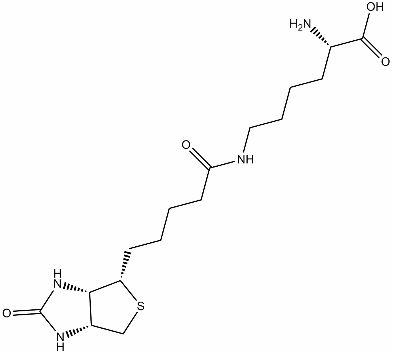
Biocytin is a compound formed by the linkage of D-biotin and L-lysine via a secondary amide bond. Biocytin serves as a classic neuroanatomical tracer, utilizing its high-affinity binding to avidin for precise localization of neuronal morphology and connectivity.
Products are for research use only. Not for human use. We do not sell to patients.
Description
Biocytin is a compound formed by the linkage of D-biotin and L-lysine via a secondary amide bond. Biocytin serves as a classic neuroanatomical tracer, utilizing its high-affinity binding to avidin for precise localization of neuronal morphology and connectivity. Biocytin is primarily used in neural circuit mapping, biotinidase activity assays, and related diagnostic research[1-2].
References:
[1] McDonald AJ. Neuroanatomical labeling with biocytin: a review. Neuroreport. 1992 Oct;3(10):821-7.
[2] Ebrahim H, Dakshinamurti K. Determination of biocytin. Anal Biochem. 1987 May 1;162(2):319-24.
Protocol
Biocytin Whole-Cell Recording and Tracing Method
This protocol is adapted from research data and provided for reference only. Adjustments may be necessary based on specific experimental requirements.
(1) Reagent and Electrode Preparation: Prepare intracellular solution containing 0.1% biocytin (components: 110mM K-gluconate, 10mM NaCl, 1mM MgCl₂, 10mM EGTA, 40mM HEPES, 2mM Mg-ATP, 0.3mM Na-GTP, pH=7.3, osmolarity 280-300mOsm). Control glass electrode resistance according to target neuron size (medium spiny neurons: 6-8MΩ; pyramidal neurons/parvalbumin interneurons: 5-6MΩ), ensuring the electrode tip is free of bubbles.
(2) Brain Slice Preparation and Neuron Localization: Obtain brain tissue from 3-month-old mice and prepare coronal slices containing the primary motor cortex (M1) or striatum (320μm thick) using a vibratome. After recovering in oxygenated artificial cerebrospinal fluid (aCSF) for 1 hour, transfer slices to a thermostatically controlled perfusion recording chamber (30°C). Identify neuronal morphology using infrared differential interference contrast (IR-DIC) microscopy (pyramidal neurons are teardrop-shaped, medium spiny neurons are oval-shaped).
(3) Whole-Cell Recording and Biocytin Loading: After the electrode contacts the cell membrane, apply negative pressure to form a gigaohm seal, then briefly apply strong suction to rupture the membrane and establish whole-cell mode. Record action potentials (AP) in current-clamp mode, then maintain whole-cell mode for 40-60 minutes to allow biocytin to fully diffuse into fine neuronal structures such as dendrites and axons. Slowly withdraw the electrode to allow the membrane to reseal.
(4) Tissue Fixation and Fluorescence Staining: Fix the recorded brain slices in 4% paraformaldehyde at 4°C for 24 hours. After permeabilization with 1% Triton-X100, incubate with streptavidin-Alexa 594 (red) or Alexa 488 (green) fluorescent probes (diluted 1:1000) at 4°C for 24 hours.
(5) Morphological Imaging and Analysis: Use a confocal microscope (63×oil immersion objective) to acquire Z-stack images of neurons (step size 0.2μm) under corresponding fluorescence signals. Perform 3D reconstruction using ImageJ software to analyze total dendritic length, branching complexity (Sholl analysis), and dendritic spine density (number of spines per unit length of dendrite).
Precautions:
(1) Experiments must comply with animal ethics guidelines.
(2) For your safety and health, please wear a lab coat and disposable gloves during operation.
References:
Tan S, Mo X, Qin H, et al. Biocytin-Labeling in Whole-Cell Recording: Electrophysiological and Morphological Properties of Pyramidal Neurons in CYLD-Deficient Mice. Molecules. 2023 May 15;28(10):4092.

You must be logged in to post a comment.